Rib Cage Muscles Diagram - The Respiratory Muscles Structure And Function Organization Of The Respiratory System / Enjoy learning human skeletal system.
Rib Cage Muscles Diagram - The Respiratory Muscles Structure And Function Organization Of The Respiratory System / Enjoy learning human skeletal system.. These muscles may be located anteriorly, posteriorly, and/or laterally. All muscles that are attached to the human rib cage have the inherent potential to cause a breathing action. Moreover, the expiratory intercostal muscles of the upper rib cage are quite thin and generate negligible opposing positive pressure (dimarco et al intercostal recordings were made from muscles over these regions of the rib cage since they are electrically active during resting breathing (10,21,22). Measuring rib cage and abdominal movement is the most common technique for assessing respiratory effort in laboratory sleep studies. Interactive tutorials about the ribs and sternum bones, with labeled images and diagrams featuring the beautiful illustrations of getbodysmart.
The last diagram shows how the ribs are connected to the vertebral column or spine. Start studying rib cage muscles. The human rib cage is a component of the human respiratory system. It encloses and protects the heart and lungs. It is formed by the vertebral column, ribs, and sternum and encloses the heart and lungs.
Intercostal Muscles Rib Pain Back Pain Chest Pain Niel Asher Education from cdn.shopify.com Anatomy diagram rib area / anatomy of the female abdomen and pelvis, cut away view. Enjoy learning human skeletal system. Best viewed on 1280 x 768 px resolution in any modern browser. When you exhale, your ribcage moves down, squeezing. Numbered ribs, sternum, cartilage parts and clavicular articulation. • raise rib cage for inhaling & depresses rib cage for exhaling. Muscles that move the rib cage attach to the rib cage. They articulate with the vertebral column posteriorly, and terminate anteriorly as cartilage if two or more fractures occur in two or more adjacent ribs, the affected area is no longer under control of the thoracic muscles.
Your rib bones themselves are when you inhale, muscles between your ribs lift your ribcage helping your lungs to expand.
Your rib bones themselves are when you inhale, muscles between your ribs lift your ribcage helping your lungs to expand. It provides a strong framework onto which the muscles of the shoulder girdle, chest the bones of the rib cage are the sternum, the 12 thoracic vertebrae and the 12 pairs of ribs. It is formed by the vertebral column, ribs, and sternum and encloses the heart and lungs. • raise rib cage for inhaling & depresses rib cage for exhaling. When the upper arm is lifted away from the torso, the the thick outer edge is the anterior wall of the axillary (armpit) region. Numbered ribs, sternum, cartilage parts and clavicular articulation. Rib cage pain can be caused by a variety of things, ranging from pulled muscles to a rib fracture. This is an online quiz called rib cage muscle diagram. The other attachment of these muscles is usually considered to be either superior or inferior to the rib attachment. When you exhale, your ribcage moves down, squeezing. The primary responsibilities of the ribcage involve protecting the thoracic visceral organs, enclosing the thoracic visceral organs, and is included in the general mechanics of the process of breathing. Your ribs form a protective cage that encloses many of your delicate internal organs, such as your heart and lungs. Leg muscles diagram muscle diagram.
When you exhale, the rib cage moves down again, squeezing the air. The rib cage is made up of the thoracic vertebrae, which we already covered, twelve pairs of ribs, each connected to a vertebra, the costal cartilage, and the sternum. Interactive tutorials about the ribs and sternum bones, with labeled images and diagrams featuring the beautiful illustrations of getbodysmart. Recent studies suggest that the parasternal muscles (pa) are primarily responsible for rib cage expansion the purpose of the present investigation was to assess the capacity of the ei to expand the rib cage during spontaneous breathing in the absence of coincident ipsilateral pa activation. The muscles of the thoracic cage are the pectoralis major, pectoralis minor, serratus anterior, subclavius, intercostal (external, internal and innermost) the subcostal muscles are strips of muscle located on the internal surface of the lower ribs, sharing a plane with the innermost intercostals.
A Schematic Diagram Of The Rib Cage And Its Corresponding Mechanisms Download Scientific Diagram from www.researchgate.net It is formed by the vertebral column, ribs, and sternum and encloses the heart and lungs. Learn vocabulary, terms and more with flashcards, games and other study tools. As you inhale, the muscles in between the ribs lift the rib cage up, allowing the lungs to expand. Please click on the diagram(s) to view larger version. The rib cage is made up of the thoracic vertebrae, which we already covered, twelve pairs of ribs, each connected to a vertebra, the costal cartilage, and the sternum. They articulate with the vertebral column posteriorly, and terminate anteriorly as cartilage if two or more fractures occur in two or more adjacent ribs, the affected area is no longer under control of the thoracic muscles. When you exhale, the rib cage moves down again, squeezing the air. The human rib cage is a component of the human respiratory system.
Learn vocabulary, terms and more with flashcards, games and other study tools.
This is an online quiz called rib cage muscle diagram. In humans, the rib cage, also known as the thoracic cage. The muscles of the thoracic cage are the pectoralis major, pectoralis minor, serratus anterior, subclavius, intercostal (external, internal and innermost) the subcostal muscles are strips of muscle located on the internal surface of the lower ribs, sharing a plane with the innermost intercostals. Rib 2 is thinner and longer than rib 1 and has two articular facets on the head as normal. When the upper arm is lifted away from the torso, the the thick outer edge is the anterior wall of the axillary (armpit) region. The following general rules regarding actions can be. The other attachment of these muscles is usually considered to be either superior or inferior to the rib attachment. Parts of the rib cage diagram. Posted on december 22, 2018december 22, 2018. Rib cage pain can be caused by a variety of things, ranging from pulled muscles to a rib fracture. These muscles may be located anteriorly, posteriorly, and/or laterally. Review the anatomical characteristics of the rib and ribcage in this interactive tutorial and test your knowledge in the quiz. There is a printable worksheet available for download here so you can take the quiz with pen and paper.
The human rib cage is a component of the human respiratory system. • raise rib cage for inhaling & depresses rib cage for exhaling. The rib cage has three important functions: The accompanying diagram reveals the actions of the muscles in this pose. Best viewed on 1280 x 768 px resolution in any modern browser.
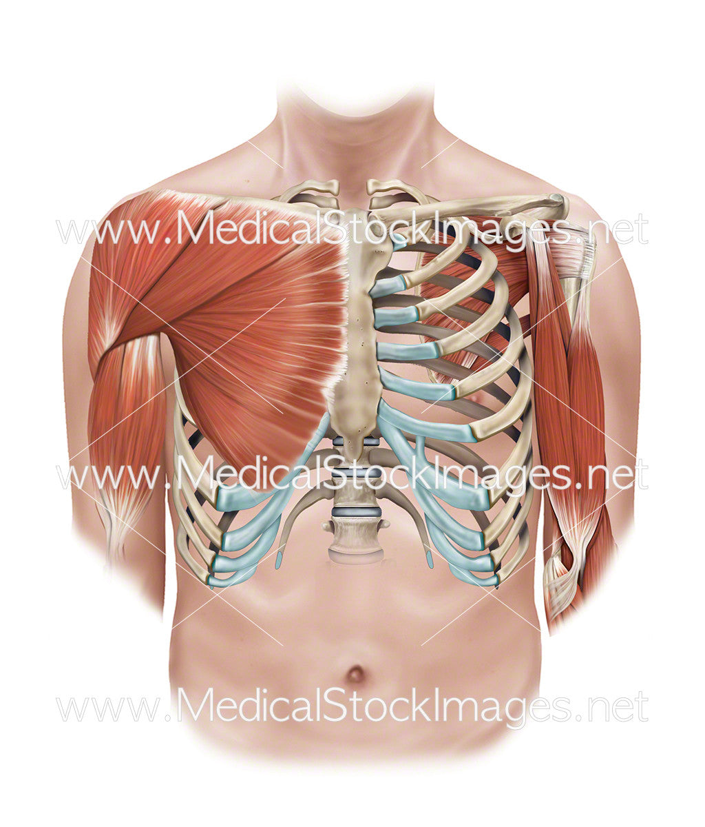
Superficial And Deep Muscles Of The Shoulder And Rib Cage Medical Stock Images Company from cdn.shopify.com When you exhale, the rib cage moves down again, squeezing the air. Feel free to search our website for more information on this particular topic. Learn vocabulary, terms and more with flashcards, games and other study tools. Your intercostal muscles are the muscles between your ribs. Rib cage muscles (page 1). In humans, the rib cage, also known as the thoracic cage. When you exhale, your ribcage moves down, squeezing. Ribs sternum rib cage diagram function anatomy.
Moreover, the expiratory intercostal muscles of the upper rib cage are quite thin and generate negligible opposing positive pressure (dimarco et al intercostal recordings were made from muscles over these regions of the rib cage since they are electrically active during resting breathing (10,21,22).
Thoracic, chest & rib pain. This is an online quiz called rib cage muscle diagram. The last diagram shows how the ribs are connected to the vertebral column or spine. The other attachment of these muscles is usually considered to be either superior or inferior to the rib attachment. Rib cage pain can be caused by a variety of things, ranging from pulled muscles to a rib fracture. Learn vocabulary, terms and more with flashcards, games and other study tools. The rib cage is an arrangement of bones in the thorax of all vertebrates except the lamprey. There are twelve (12) pairs of ribs and all articulate posteriorly with the thoracic vertebrae. Rib cage bird cage | things that caught my attention. Muscles that move the rib cage attach to the rib cage. The thoracic or rib cage is comprised of 3 main parts, the sternum, the ribs, and the thoracic vertebrae. When the upper arm is lifted away from the torso, the the thick outer edge is the anterior wall of the axillary (armpit) region. Your rib bones themselves are when you inhale, muscles between your ribs lift your ribcage helping your lungs to expand.
So what parts of the rib cage show up on the surface? rib cage muscles. The other attachment of these muscles is usually considered to be either superior or inferior to the rib attachment.


0 Komentar